|
Automatic Detection and Measurement of Structures in Fetal Head Ultrasound Volumes Using Sequential Estimation and Integrated Detection Network (IDN)
Abstract
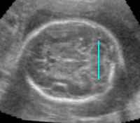
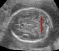
Routine ultrasound exam in the second and third
trimesters of pregnancy involves manually measuring fetal head
and brain structures in 2D scans. The procedure requires a
sonographer to find the standardized visualization planes with
a probe and manually place measurement calipers on the
structures of interest. The process is tedious, time consuming,
and introduces user variability into the measurements. This
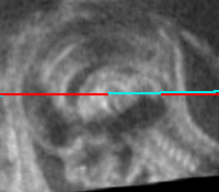
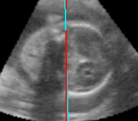
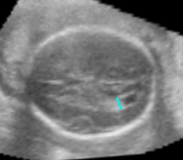
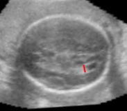
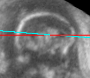
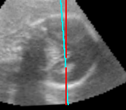
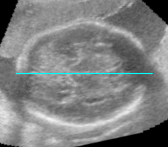
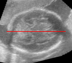
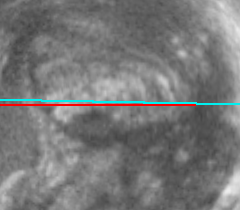
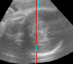
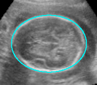
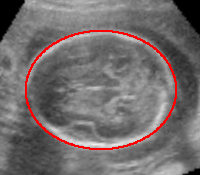
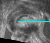
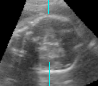
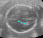
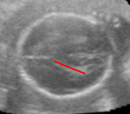
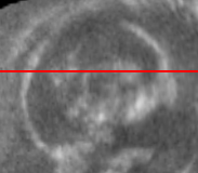
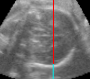
paper proposes an Automatic Fetal Head and Brain (AFHB) system
for automatically measuring anatomical structures from 3D
ultrasound volumes. The system searches the 3D volume in a
hierarchy of resolutions and by focusing on regions that are
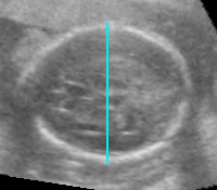
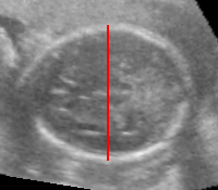
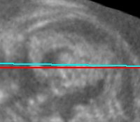
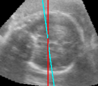
likely to be the measured anatomy. The output is a standardized
visualization of the plane with correct orientation and
centering as well as the biometric measurement of the anatomy.
The system is based on a novel framework for detecting multiple
structures in 3D volumes. Since a joint model is difficult to
obtain in most practical situations, the structures are
detected in a sequence, one-byone. The detection relies on
Sequential Estimation techniques, frequently applied to visual
tracking. The interdependence of structure poses and strong
prior information embedded in our domain yields faster and more
accurate results than detecting the objects individually. The
posterior distribution of the structure pose is approximated at
each step by sequential Monte Carlo. The samples are propagated
within the sequence across multiple structures and hierarchical
levels. The probabilistic model helps solve many challenges
present in the ultrasound images of the fetus such as speckle
noise, signal drop-out, shadows caused by bones, and appearance
variations caused by the differences in the fetus gestational
age. This is possible by discriminative learning on an
extensive database of scans comprising more than two thousand
volumes and more than thirteen thousand annotations. The
average difference between ground truth and automatic measu-
ements is below 2 mm with a running time of 6.9 seconds (GPU)
or 14.7 seconds (CPU). The accuracy of the AFHB system is
within inter-user variability and the running time is fast,
which meets the requirements for clinical use.
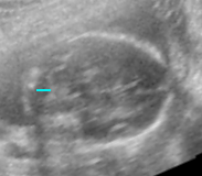
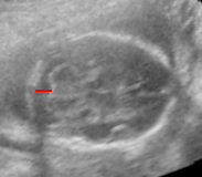
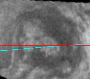
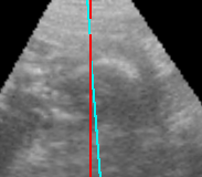
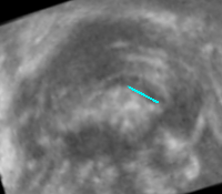
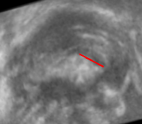
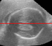
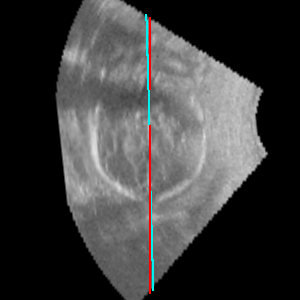
Results
Publications and Further Reading
|
Automatic Detection and Measurement of Structures in Fetal Head Ultrasound Volumes Using Sequential
Estimation and Integrated Detection Network (IDN)
Michal Sofka and Jingdan Zhang and Sara Good
and S. Kevin Zhou and Dorin Comaniciu
IEEE Transactions on Medical Imaging (TMI), vol. 33, no. 5, pp. 1054-1070, May 2014.
[pdf]
[bibtex]
[publisher]
|
|